Magnetic resonance imaging (MRI) shows changes in bone and cartilage and can discriminate these from fluid and soft tissue around the joints, making it a good technique to measure synovial volume and inflammation characteristic of rheumatoid arthritis (RA).
What is a pannus in rheumatoid arthritis?
Pannus is a type of extra growth in your joints that can cause pain, swelling, and damage to your bones, cartilage, and other tissue. It most often results from rheumatoid arthritis, an inflammatory disease that affects your joints, though other inflammatory diseases are also sometimes to blame.
Why would a rheumatologist order an MRI?
Basic indications for MRI examinations in rheumatoid patients include [1–3]: assessment of inflammatory lesions of joint cavities, sheaths and bursae (synovial thickening, synovial congestion, effusion); assessment of bone lesions (bone marrow edema, geodes, erosions, damaged articular cartilage);
Does rheumatoid arthritis affect subchondral bone?
It is characterised by persistent synovitis and formation of pannus, i.e. invasive synovial tissue, which ultimately leads to destruction of the cartilage, subchondral bone, and soft tissues of the affected joint.
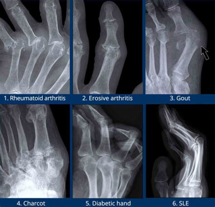
Can you be misdiagnosed with rheumatoid arthritis?
Misdiagnosis of Rheumatoid Arthritis Is Common. Many rheumatic diseases have overlapping symptoms, and that can complicate the effort to obtain an accurate diagnosis for RA. Some diseases are complex. They may have overlapping characteristics with other conditions, making diagnosis more difficult.
Does RA cause brain fog?
RA causes chronic inflammation. Along with its effects on the joints, RA may cause brain fog, which can involve difficulty concentrating, poor memory, or confused thoughts.
How is pannus formation in rheumatoid arthritis?
Rheumatoid arthritis causes the immune system to attack the synovium. The synovium then becomes inflamed, forming pannus. This process typically happens gradually, causing symptoms to develop over weeks or months.
Can you see muscle inflammation on an MRI?
MRI is sensitive in detecting muscle inflammation, but it is not specific to a diagnosis of myositis because muscular dystrophies and other myopathies may have associated edema on MRI [2]. The signal changes on imaging need to be interpreted in the context of the clinical setting.
Can RA cause numbness?
RA sometimes affects the small nerves in your hands or feet. They might feel numb or like you’re being stuck with pins and needles. If these tiny blood vessels in your hands or feet shut down, your fingers or toes may feel cold or numb. They could even change color when it’s cold outside and look white, red, or blue.
What autoimmune mimics rheumatoid arthritis?
Lupus and Scleroderma The autoimmune diseases systemic lupus erythematosus and scleroderma often present with joint involvement that mimics rheumatoid arthritis. While lupus and scleroderma are two different diseases, they often overlap with one another.
How can MR imaging help diagnose rheumatoid arthritis (RA)?
List the MR imaging features that can help establish an early diagnosis of rheumatoid arthritis. •. Identify the MR imaging findings of enthesitis that help differentiate rheumatoid arthritis from some clinical subsets of peripheral spondyloarthropathies.
What is pannus in rheumatoid arthritis (RA)?
Loading images… Large soft tissue mass encircling the eroded dens consistent with “pannus”, i.e. hypertrophied synovium. Minor extrinsic compression to the upper cervical cord. Marked erosion of the dens and adjacent joints with pannus (soft tissue mass) is typical of advanced rheumatoid arthritis.
Which physical findings are characteristic of advanced rheumatoid arthritis (RA)?
Marked erosion of the dens and adjacent joints with pannus (soft tissue mass) is typical of advanced rheumatoid arthritis.
What is MRI of synovitis in rheumatoid arthritis?
MRI OF SYNOVITIS IN RHEUMATOID ARTHRITIS. The phenomenon of contrast enhancement seen in RA synovitis occurs very quickly on intravenous administration of a contrast agent and thus reflects an unusually rapid increase in the concentration of gadolinium that occurs within the abnormal (but not within normal) synovium.
