A standard ultrasound imaging test won’t definitively tell your doctor whether you have endometriosis, but it can identify cysts associated with endometriosis (endometriomas).
What imaging shows endometriosis?
Currently, MRI is considered the best imaging technique for mapping endometriosis, since it provides a more reliable map of deep infiltrating endometriosis than physical examination and transvaginal ultrasound (TVUS) [8].
How is deep infiltrating endometriosis diagnosed?
Imaging studies and invasive diagnostic tests
- Magnetic resonance imaging (better than CT) is the method of choice when adenomyosis or deep infiltrating endometriosis is suspected.
- Intravenous urography (if ureteric involvement is suspected)
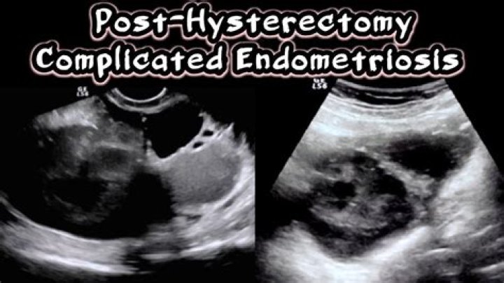
What cysts are associated with endometriosis?
Pathological cysts occur when endometriosis affects the ovaries. Endometriotic tissue can either grow on the surface on the ovary or deep inside it. Deep ovarian endometriosis is known as endometriomas or ovarian cysts. It causes the formation of cavities within the ovary that fill with blood.
Can Endometrioma be seen on ultrasound?
What your doctor will look for on an ultrasound scan. Your doctor will look for endometrioma — a type of ovarian cyst — on an ultrasound scan for diagnosing endometriosis. If you have endometrioma on the scan, your doctor may use that for diagnosis or order other scans and tests to confirm it.
Can an abdominal CT scan detect endometriosis?
Plain film radiography, computed tomography (CT) scanning, and barium studies are not sensitive for the diagnosis of endometriosis. Moreover, the appearance of implants and endometriomas is nonspecific. US scanning and MRI are not sensitive for superficial lesions.
Can endometriosis be seen on imaging?
Your doctor has ordered an MRI (magnetic resonance imaging) exam to detect sites of deep endometriosis in the pelvis, which can help with surgical planning. Once these sites are surgically removed, you should have fewer symptoms.
What is abdominal wall endometriosis?
Abdominal wall endometriosis (AWE) is defined as implantation of endometrial tissue outside the peritoneum, including lesions secondary to a surgical incision and those that arise spontaneously [1].
What organs are affected by endometriosis?
Generally, endometriosis is found in the pelvic cavity. It can attach to any of the female reproductive organs including, but not limited to, the outside of the uterus, fallopian tubes, ovaries, uterosacral ligaments, peritoneum, and any of the spaces between the bladder, uterus, and vagina.
Does ultrasonography have a role in the evaluation of abdominal aortic aneurysm?
The aim of the study was the assessment of the diagnostic value of computed tomography and ultrasonography in the evaluation of abdominal aortic aneurysm. Material comprises a group of 26 patients with abdominal aortic aneurysm. There were 18 men and 8 women, aged between 48 and 76 years (mean age 62 years).
What are abdominal aortic aneurysms?
Abdominal aortic aneurysms (AAA) are focal dilatations of the abdominal aorta that are 50% greater than the proximal normal segment or >3 cm in maximum diameter.
What does an enlarged area in the lower part of aorta mean?
The enlarged area in the lower part of the aorta is an abdominal aortic aneurysm. An ultrasound image of an abdominal aortic aneurysm is shown in the upper right corner. Ultrasound imaging is often used to diagnose abdominal aortic aneurysms.
How is an aneurysm diagnosed in a lumbar spine?
An aneurysm may be visible as an area of curvilinear calcification in the paravertebral region on either abdominal or lumbar spine radiographs performed for alternative indications. Ultrasound assessment is simple, safe and inexpensive.
